This week has been the busiest by far for me. Since I'll be out of town next week, I have ensured that all of my duties have been performed in such a way to last an extra week. Western blots, cardiomyocyte isolations, buffer preparation, lipid lesion analysis, and surgeries on our specimen are still running rampant in the lab.
After watching my DNA Gel Electroporesis video, Dr. McConnell, my PI, suggested that I create a library of lab videos to show incoming students as well as students who are undergoing their rotations within the department the proper technique for basic lab functions.
Storing Western Blot Membranes-Lauren Tolat from Lauren Tolat on Vimeo.
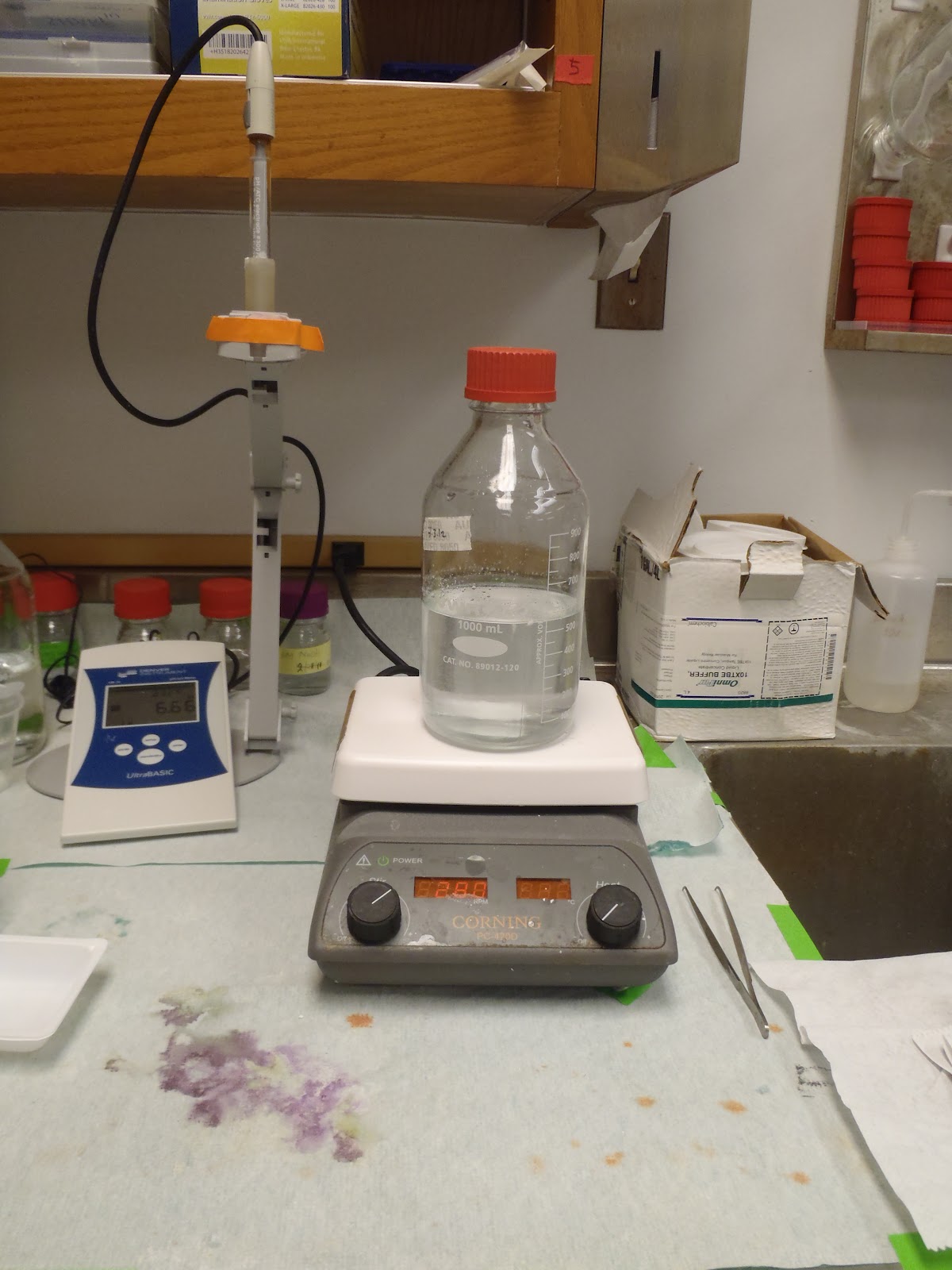
How to: Standardize and Use the pH Meter - Lauren Tolat from Lauren Tolat on Vimeo.
The first video demonstrates the technique (and some tips) involved in storing Western Blot membranes. A common way to store the membranes is by transferring them to a polyethylene bag and keeping them saturated in TBST. The final product is stored in the refrigerator (2 to 8 degrees Celsius).
The second video is a technique used in general lab settings-standardizing and using the pH meter. When preparing a high volume of buffer solutions that need specific pH and temperature adjustments, the pH meter is a lifesaver. However, many lab students are unaware of the lengthy procedure involved in standardizing and cautiously using the meter for their own measurements.
Overall, I am beyond ecstatic about ALL of the opportunities that I have been able to experience during my undergraduate research and I feel incredibly blessed to be here at the University of Houston.
Friday, July 27, 2012
Friday, July 20, 2012
Week 9-DNA Gel Electrophoresis
This week has been super busy! Among cardiomyocyte isolations, western blots, protein assays, operations on our live specimen, and a potluck party thrown in honor of my graduate student mentor's successful research proposal, I had the opportunity to perform my very own DNA Gel Electrophoresis. Electrophoresis is a method used in this lab for genotyping among our six different experimental groups. Specifically, it is used to determine whether our gene of interest is available in a particular sample after PCR has been performed. It involves four steps: 1) preparing the gel; 2) loading the gel; 3) running the gel; and, 4) visualizing. With the lovely help of my multi-talented research team, I created a video of me performing these steps.
 |
| An Assortment of Food-Potluck Party |
 |
| The Team-Potluck Party |
 |
| The Team-Potluck Party |
 |
| Cori and Fan |
 |
| Western Blots |
 |
| An Alternative to Milk in Western Blotting-BSA |
 |
| Lauren's Homemade Stripping Buffer :) |
 |
| Casket used to Mold Gel |
 |
| Running the Gel |
 |
| Protein Electrophoresis-Loading Samples |
 |
| Checking the Current |
 |
| Light Cabinet-Visualizing |
 |
| Light Cabinet-Visualizing |
 |
| Software used in Electrophoresis |
 |
| Visualizing the Bands |
 |
| Choosing a Filter to View the Results of Electrophoresis |
 |
| Transferring Membranes to Polyethylene Bags |
 |
| Transferring Membranes |
 |
| Storing Membranes |
 |
| Preparing Hypotonic Buffer for Cell Culture |
Friday, July 13, 2012
Week 8-Solutions, Buffers, and the Joy of Scientific Replication
Congratulations are in order for Ms. Qiying Fan, the hard-working and dedicated Ph.D. graduate student, who successfully completed her graduate research proposal this week. The entire research team is so proud of her and we couldn't have asked for a more positive response from the committee.
The entire research team and our PI have been pretty busy-writing grants and conducting/planning experiments.
Andrea has been planning experiments and the next steps for her project which is likely to span months. During the previous weeks, she had been refining her skills at inducing myocardial infarctions on her live specimen (mice). Now, with her technique finalized, she is officially ready to begin the actual experimental part of the project. She will be injecting certain stem cells into her specimen to test whether these cells improve cardiac function after the infarction. Her overreaching goal is to figure out if these stems cells aid in the recovery of localized damaged tissue in the heart.
Abeer has been continuing her experiments involving cardiomyocyte isolations. Once she obtains the cardiomyocytes (the cells that comprise cardiac muscle), she performs measurements to figure out the calcium ratio present using a specialized miscroscope called the Ionoptix. The calcium ratio will determine the degree of contractility in the cardiomyocytes.
Fan, while still recovering from a stressful week, has been continuing the development of Western blots to detect different treatments of Bapta-AM, a calcium inhibitor (used because calcium is involved in the specific pathway she is trying to identify). She has been treating VSMC (vascular smooth muscle cells) to see how specific growth factors affect VSMC intracellular signaling. Overall, this will further her understanding of how calcium, and how much with her different experimental groups, is involved in the pathway.
I had to opportunity to make my own Western Blot Stripping Buffer using substrates from the lab. A stripping buffer is used in Western blots before the blocking stage to remove primary and secondary antibodies from the PVDF membrane and allow chemiluminescent Western blots to be reprobed. This saves time and is more efficient than continuously performing gel electrophoresis and duplicate immunoblot assays. In other words, it allows the experimenter to reuse a PVDF blot to detect a different target with a different primary antibody.


 |
| Congrats to Fan! |
 |
| Tony Ohonba-Pharmacy Student |
 |
| My Graduate Student Mentor and I |
Abeer has been continuing her experiments involving cardiomyocyte isolations. Once she obtains the cardiomyocytes (the cells that comprise cardiac muscle), she performs measurements to figure out the calcium ratio present using a specialized miscroscope called the Ionoptix. The calcium ratio will determine the degree of contractility in the cardiomyocytes.
Fan, while still recovering from a stressful week, has been continuing the development of Western blots to detect different treatments of Bapta-AM, a calcium inhibitor (used because calcium is involved in the specific pathway she is trying to identify). She has been treating VSMC (vascular smooth muscle cells) to see how specific growth factors affect VSMC intracellular signaling. Overall, this will further her understanding of how calcium, and how much with her different experimental groups, is involved in the pathway.
I had to opportunity to make my own Western Blot Stripping Buffer using substrates from the lab. A stripping buffer is used in Western blots before the blocking stage to remove primary and secondary antibodies from the PVDF membrane and allow chemiluminescent Western blots to be reprobed. This saves time and is more efficient than continuously performing gel electrophoresis and duplicate immunoblot assays. In other words, it allows the experimenter to reuse a PVDF blot to detect a different target with a different primary antibody.
 |
| Effect of Stripping Buffer on Membranes |
 |
| Mild Stripping Buffer-Protocol |

 |
| Glycine-15 g |

 |
| SDS-1 g |
 |
| Tween 20-10 mL |
 |
| Adjusting the pH to an Acidic Level |
 |
| Adjusting pH |
 |
| The Finished Product |
 |
| Aluminum Foil used to Protect Against Light Sensitivity |
 |
| Commercial Stripping Buffer |
So, instead of spending $134 to buy a commercial buffer, why not recreate it on your own? :)
Other activities this week included preparing transfer buffer, running buffer, developing Western blots, transferring tissue to histology cassettes, and autoclaving.
Friday, July 6, 2012
Week 7-The Research Proposal
I hope everyone had a wonderful Independence Day!
One thing that Fan's research presentation showed me was the importance of having a strong connection between the background and the preliminary data. The background indicates the significance of the problem to be examined in the research project while the preliminary data targets a specific aspect of the problem. In this specific situation, Fan needed to make a stronger connection between her notes about atherosclerosis and the protein that she is investigating in the signal transduction pathway.
Additionally, two graduate students from TSU have been touring the facility lately. Dr. McConnell said that they are prospective lab members and are in the process of learning specific lab protocols/techniques. I was asked to introduce these students to what I've learned over the course of my stay here in the lab, including: autoclaving, filtration, and Western blots. How exciting is that? Just a few weeks ago I was in the process of being trained, and now, I get the opportunity to instruct. It's such a rewarding feeling.
This week I participated in developing Western blots, transferring heart tissue into histology cassettes, and preparing buffers for cardiomyocyte isolation. One buffer I had a fun time making was the Fura 2-AM loading buffer (cardiomyocyte perfusion buffer). It was difficult making this buffer because the substrates do not fully dissolve in the solution unless the pH is adjusted. The contents make the solution basic so hydrochloric acid needs to be added in order to reach proper homogeneity. Fura 2-AM is an intracellular calcium indicator that is ratiometric and UV light excitable. At low concentrations of the indicator, accurate measurements of the intracellular Ca2+ concentration are able to be measured.
 |
| Cardiomyocyte Buffer-Preparation Sheet |
 |
| Cardiomyocyte Buffer-Preparation |
 |
| Sonal Performing Cardiomyocyte Isolation |
 |
| Solution-Making |
 |
| Solution-Making |
 |
| Cori and I |
 |
| Solution Making |
 |
| Adjusting the pH |
 |
| Cori :) |
 |
| The Finished and Filtered Buffer! |
 |
| Fan Preparing Her Research Proposal |
 |
| Fan-Practicing Her Presentation |
Subscribe to:
Posts (Atom)